


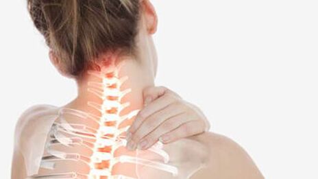
Cervical osteochondrose is a progressive degenerative-dystrophic process, which leads to exhaustion, deformation and destruction of intervertebral discs of the Cervical region.The loss of shock - the magical cartilage causes pain and due to the exposure of articulated surfaces (spondylarthrosis), and due to the movement of the nerves of the root of the spinal cord.
In the absence of timely treatment, it is possible to mitigate spine with the loss of its natural flexibility, the injured blood supply, deterioration of nervous enforcement in those parts of the body that find roots of cervix spine.
Pathology can also develop both independently and as part of the generation of spine with covering breasts, lumbar and sacral parts.
General information
It is believed that osteochondrosis of the neck spine is more common than in other departments.In fact, this is not so - dystrophic phenomena develop evenly at all maximum load points - in the field of the main bend of the spinal column (the lower part is located, it is a greater load that carries).However, the symptoms of cervical osteochondrosis are more pronounced, so they look more often.It is due to the high mobility of the vertebral door, which holds the head at the same time, as well as with the trait of the root of the spinal cord.
Note!According to statistics, the disease affects more than 60% of the middle and older people.However, recently, the rejuvenation of the pathological process has been observed - pathology is in young people, and even in adolescents.This is due to the general computerization of studies and work, as well as reducing physical activity and deterioration of the quality of the diet.
Given the age audience, 2 forms of cervical osteochondrone - physiological and pathological are different.
Physiological processIt is related to the natural aging of the body, when the symptoms of the disease are due to the gradual spending of intervertebral disks.The process arises under the influence of the endocrine system and is the consequence of the menopause.The destruction of the cartilage structure begins from the center of the intervertebral disk and accompanied by gradual replacement of cartilage tissue fibrous.The pathology is irreversible, however, can be compensated with special medicines.
Pathological processIt is related to abnormal destructive changes in the body - immune, dystrophic, inflammatory, metabolic.First of all, subcutaneous tissues are involved salt salts on bone structures, nerve roots are inflamed, atrophy or skeleton muscle hyperstonity, leading to circulatory disorders in their heads and grooves.With timely diagnosis, pathology is treated and completed with complete renewal of a healthy function of organ and tissue.
Stages of cervical osteochondrosis and their symptoms
There are 4 main phases of the pathological process:
- 1. phase - expresses slight discomfort and muscular overpatement in the sick region, Hartilege disks lose stability;
- Another phase-local pain appears, especially with head movements.Intervertebral discs are deformed, the fibrous ring begins to assemble, the distance between vertebral is reduced;
- 3. Phase - The pain is amplified and becomes constant, movement becomes limited.Chiefs of their heads can cause stain, nausea, brain blood breaches to general fatigue, cartilage tissue, the vertebrae are closed, the brainstorm is completely destroyed with the emergence of the risk of intervertebral hernia;
- Fourth phase - The pain syndrome completely immobilizes the area of the door, the bloodshed blood of the brain is disturbed and requires constant medication support, vertebrates begin to grow together.
Cervical osteochondrosis: Signs, symptoms of pathology
In the first phases of osteochondrosis is asymptomatic.As the disease develops, a characteristic characteristic is the presence of painful or unpleasant sensations in the head, neck and chest, less often the upper limbs.
All possible symptoms can be conditionally attributed to 4 types of syndrome: heart, vertebrae, record (nerve) and spinal artery syndrome (with circulatory disorders).
Vertebra syndrome:
- crumb in neck when turning / tilting heads;
- With the progress of the disease, pain and difficulty movement occur;
- Morphological disorders of the structure in the body of the vertebrae and intervertebral space (visible on X -Ray).
Heart syndrome:
- shortness of breath, weakness;
- A sense of incomplete breath, lack of air;
- Spontaneous phenomena from cardiovascular system-angina pectoris, pain cloths, ignition;
Rook syndrome:
- Tongue and arms, fingers, occipital regions;
- difficulty swallowing;
- an unpleasant sensation in the area between the spatula;
- Headache in order and forehead.
Vail Artery syndrome:
- Unreasonable jumps in blood pressure;
- dizziness, to loss of consciousness;
- noise in ears, a feeling of cotton wool in the head;
- A temporary one-treaded blindness, "flies" in the eyes;
- Periodic attacks nausea, especially when title;
- Headaches - mostly in napes, as well as migraines;
- Drowsiness, performance reduction, memory, attention concentrations, depression.
Attention!All these syndromes should be combined with each other.The absence of symptoms of one of them may be the reason for the reason differential diagnosis with other disease groups.
Cervical osteochondrose causes
Dorrupt phenomena in the cervical spine are connected to the vertical location of skeletons and the specific distribution of static and dynamic loads, which is largely dependent on the prevailing posage and the degree of skeletal muscle development.
Main reasons:
- Lack of movement - what is not developing - degradates: weak muscles, tissues were destroyed;
- Incorrect static poses - muscle clamps lead to circulatory disorders with subsequent tissue dystrophy;
- Lack of nutrition or unbalanced diet - The body should be received all necessary for the construction and renovation of bones and cartilage structures of the skeleton, maintaining a muscle tone;
- Obesity, excess weight, weight wear - the load on the skeleton structure increases;
- constant nerve tension and nerve voltages;
- The hypothermia of the cervical region - "caught", "inflated" - causes hidden inflammatory processes;
- The presence of autoimmune diseases with involving cartilage tissue leads to its premature destruction;
- Endocrine pathologies are confused with mineral metabolism, reduce the digestibility of calcium, silicone, phosphorus and other elements of bone tissue-cristotine;
- violations of the cervical region;
- Congenital abnormalities of spine and adjacent muscles.
Diagnostics
The diagnosis of "osteochondrose cervical vertebrae" consists of a low specificity of symptoms and a wide range of their events.The testing process will require a consultation of neurologists, surgeons, orthopedists, cardiologists.
Physical examination performs a doctor with a patient survey.The main diagnostic load lies in instrumental and laboratory research methods.
Instrumental diagnostics:
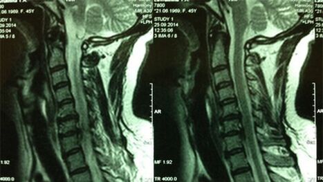
- X -ray of cerrvical sector;In the initial phase of the procedure, the MRI by the Cervical Department will be informative - will provide high visualization of solid and soft tissues - the situation of interfood drives, the presence of osteophytes, deformation, nervous roots and blood vessels;Will assess the state of ligaments, muscles, bone tissues;
- Ultrasound shows the dynamic condition of soft tissues;
- Dopplerography of the vessel doors will assist in the assessment of hemodynamics and degree damage to blood vessels (especially, the state of the spinal artery);
- Contrast myelography - will help with the suspect of violation of nerve processes;
- ECG and hearing echocardiography are used for differential diagnosis of heart syndrome with cardiovascular diseases.
How to treat cervical osteochondrose
The medical measure complex is formed taking into account the phase of the disease (acute, chronic), the degree of damage and the cause of pathology development.Use conservative treatment, surgical intervention, mixed approach.
Conservative effect
It is a gradual renewal or compensation for damage to the background of symptomatic treatment.Includes drug therapy, physiotherapy, exercise therapy and massage methods.
Medicine treatment:
- With a figure laterally side side lateral side bogs and fat of local impact;in severe cases - the usual hospital in the form of tablets;
- Anti -infalmator drugs - NSAIDs, as well as corticosteroids (a short course if necessary);
- Medications to improve microcirculation and blood circulation in general;
- Chondroprotectors - Funds for protection and return of cartilage fabrics;
- Musorelaxans - to remove muscle clamps and cramps;
- Vitamin and microelen complexes are needed for nutrition and support of the fabric by building elements.
As acute symptoms are weakened, the methods of physiotherapy, exercise therapies and self-consideration are linked.
Therapeutic gymnasticsImproves the nutritional nutrition and bone tissue by returning the blood supply in the damaged area.To avoid complications, the use of isometric movements is recommended, when instead of actual turns and slopes of heads that can damage, they use their imitation.
Attention!All actions should be done only after diagnosis and consulting with a doctor.
The technique allows you to develop and strengthen the atrophied muscles of short doors and stabilize the position of the neck of the spine.Follow of basic exercises:
- The right hand palm is shortened on the side surface of the head-palm on the head within 10 seconds, while the heads of the head and the resistance door is swollen - the head should remain stationary.
- Reduce your hand, relax your head and neck muscles as much as possible in 20 seconds.
- Repeat the exercise with your left hand.
- Color both hands with your palms in the forehead led by forehead for 10 seconds, as if trying to refuse your head back, while the neck muscles are swollen.The head should remain motionless.
- Reduce your hands, relaxing muscles as similar as the previous exercise.
- Place both palm trees in the area above the back of the head.Per Analuge, make loads of load on neck muscles, trying to tilt your head forward - it should be immovable.
- Reduce your hands, relax your neck and head muscles.Repeat the set of exercises 4-10 times.
After strengthening the short door muscles, you can start performing dynamic exercises.
Note!As a methodology for restoring the mobility of the cervical regions, the gymnastics of swimming and water proved well.
Self-considerationAllows you to reduce the intensity of local reactions and remove muscle clamps during static work.Execution rules:
- Exposure area - rear of head, back and side surfaces;
- perform the procedure in a seated position;
- movements should be performed in the direction of the spine;
- Use only supreme fingers;
- Avoid pressure on inflamed surfaces;
- Make movements Smooth - sharp pressure can harm.
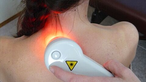
PhysiotherapyCharacteristic of rehabilitation of hospital treatment and sanatorium resorts.Well proven:
- Electrophoresis - hears the zone, improves microcirculation, used for deeper penetration of local drugs;
- Magnetotherapy;
- amplipulse;
- Uhf.
The surgical intervention is marked with complicated extrusion, breaches of spinal cord and unconscious pain syndrome.
Than cervical osteochondrose is dangerous
The area of the door concentrates the thick intertwining of the main blood vessels, nerve processes and dynamic bone bone structures.In the absence of treatment, serious pathological changes can be noticed:
- The weakening of the fibrous ring causes dislocations and subbruxation in the most modern vertebrae;
- The presence of osteophytes and spasm muscles leads to a violation of nervical roots and blood vessels with the formation of compression syndrome;
- Destruction of crispy disks and the rapprochement of vertebrae leads to intervironmental piles with a violation of nervous tissues.
Each of this phenomenon follows the pronounced negative reaction from the entire organism.
Possible complications and consequences
The list of the most common complications of cervical osteohondrone can be included:
- Vegetascular Dystonia;
- hypertension;
- Actioned smooth brain and its structures;
- Retry eye dystrophy with vision impairment;
- The failure of the tyroid failure;
- Disorder of the function of Esophagus and trachea - difficulties in swallowing and breathing with cramps;
- unconscious syndrome pain in his head, neck, chest, upper extremities;
- Convulsions and stiffness of the face, hands;
- A hypothalamic-pituitary-pituitary-pituitary disorders that draw the failure of the entire hormonal body activity.
Preventive measures
The most efficient treatment is the prevention of the disease.Prevention will help you with that.It is enough to follow several basic recommendations:
- Adjust your posture
- create a practical workplace;
- During sitting work, separate the breaks of "physical education";
- Involve in your diets of food rich in calcium, magnesium, phosphorus, silicoma - fish, nuts, seeds, legumes, dairy products, fresh vegetables, fruit;Limit the consumption of salt, sweet, flour and sharp dishes;
- To sleep and rest, use an orthopedic mattress and a pillow;
- Take care of non-average sports - it's better to give the advantage of swimming.
Even if you are unable to take into account all requirements, moderate physical effort, proper nutrition and careful relationship to your position can significantly reduce the risk of pathology.